MAXILLOFACIAL TRAUMA: CURRENT PRACTICE IN MANAGEMENT AT PAKISTAN INSTITUTE OF MEDICAL SCIENCES
S. Shahid Hussain, Muhammad Ahmad, M. Ibrahim Khan, Muhammad Anwar, Mughese Amin, Sameera Ajmal, Farhan Tariq, Naheed Ahmad, Tariq Iqbal, Saleem A. Malik
Department of Plastic Surgery, Pakistan Institute of Medical Sciences, Islamabad.
Background: This study was carried out to determine the aetiology, pattern and management of maxillofacial injuries at PIMS, Islamabad. Methods: This descriptive study was conducted at Plastic Surgery Department, PIMS Islamabad from 1st February 1998 to 30th April 2002. All the adult patients presenting with maxillofacial injures were included where as patients less than 12 years of age and only facial lacerations were excluded. Similarly isolated nasal bone fractures were also excluded because these patients were routinely managed by ENT department. Age, sex, presentation, aetiology, associated injuries and treatment modalities undertaken in these patients were recorded. RESULTS: In 164 patients 254 fractures were noted. Most were male (86%), ranging in age from 13–71 years with a male to female ratio of 6:1 respectively. The most frequent (48%) cause noticed was road traffic accidents followed by assault. Mandible was the commonest to be involved in such injuries followed by maxilla. Most of the patients (32%) had associated facial injuries. Various treatment modalities were practiced. Conclusion: Maxillofacial fractures should be managed by open reduction and internal fixation as early as possible.
Key Words: Maxillofacial, Trauma, Mandible, Management
INTRODUCTION
Maxillofacial trauma is presented in Accident and Emergency Department of the hospital as isolated injuries or part of poly trauma1. Maxillofacial trauma can be limited to superficial laceration or abrasion or it may be associated with multiple injuries to the chest, head, cervical spine, abdomen or the extremities2. It not only hampers the function but also causes serious psychological and cosmetic deficiencies2. Some of the most severe maxillofacial injuries are caused by automobile accidents but many others may result from industrial accidents, sports, home accidents and missiles or gun shots2. The frequency of facial injuries is high because face is exposed and because there is little protective covering3. A unique aspect of facial injuries is that the restoration of appearance may be the chief indication for the treatment3. The treatment of such injuries is accomplished in three phases. The primary phase deals with survival of the patient by maintenance of haemodynamics and airway function. In the intermediate phase, supportive line such as antibiotics prophylaxis and treatment of infections, control of bleeding, and tissue debridement are done. The third phase is the reconstructive phase. The aim of this phase is reconstruction of the soft and hard tissues (using grafts if required), reduction and fixation of bone segments, reconstruction of the nasolacrimal system, release of scar tissue, and correction of sensory and motor nerve dysfunction4.
Epidemiological studies of maxillofacial trauma have classically shown that young adult males are the predominant victims5. Maxillofacial injuries are not uncommon in Pakistan6. A compromised cosmetic, functional, and psychological outcome may result when these components of successful treatment are not practiced7.
This study was conducted to determine the aetiology, pattern and management of maxillofacial injuries at PIMS, Islamabad.
MATERIAL AND METHODS
This descriptive study was conducted at Plastic Surgery Department of PIMS Islamabad from 1st February 1998 to 30th April 2002. All the adult patients presenting with maxillofacial injures were included where as patients less than 12 years of age and only facial lacerations were excluded. Similarly isolated nasal bone fractures were also excluded because these patients were routinely managed by ENT department. Age, sex, presentation, aetiology, associated injuries and treatment modalities undertaken in these patients were recorded.
Almost all of the patients presented in Accident and Emergency Department. The management started with ATLS (Advanced Trauma Life Support) including the maintenance of air way, control of bleeding, antibiotic coverage and head end elevation at 45°. Barton’s bandage was applied to achieve the normal occlusion wherever possible. Regular mouth washes and liquid diet were advised. In all cases plain x-rays, i.e., OPG (orhtopantomogram), Water’s view or CT scan were obtained when possible. Majority of the patients were operated on routine lists. Inter Maxillary Fixation (IMF) was done in isolated mandibular fractures when the pre-injury occlusion was achieved easily under local anaesthesia (infraorbital and mental nerve blocks) and intra venous sedation. It was done with cortical screws and 27 Fr dental wires. It remained there for 4–6 weeks. During this period the patients remained on liquid diet.
Two or more mandibular fractures with or without flail segments, maxillary, zygomatic, orbital rim fractures were approached via intra-oral route (gingivolabial incision). Rarely only those fractures were approached from external wound when it lied on the line of fracture not closed primarily. All these patients were operated under general anaesthesia with north nasal intubation. Nasogastric intubation was done for 24 hours to avoid vomiting and accidental aspiration.
Descriptive statistics were used to calculate percentages.
RESULTS
In 164 patients, 254 fractures were noted. Most of the patients were males (86%) with a male to female ratio of 6:1 and age ranging from 13–71 years (mean 36.4 years).
The most frequent (48%) cause noticed was road traffic accidents followed by assault (Table-1).
Table-1: Aetiology of Maxillofacial Injuries (n=164)
|
Cause |
Patients |
% |
|
Road Traffic Accidents |
78 |
47.6 |
|
Assault |
49 |
29.9 |
|
Domestic Accidents |
21 |
12.8 |
|
Industrial Accidents |
9 |
5.4 |
|
Gunshot |
6 |
3.7 |
|
Sports Injuries |
1 |
0.6 |
Mandible was the commonest to be involved in such injuries followed by maxilla (Table-2).
Table-2: Areas Involved in Maxillofacial injuries
|
Areas/Bones |
Patients |
% |
|
Mandible |
132 |
62.9 |
|
Zygoma |
28 |
13.3 |
|
Maxilla |
24 |
11.4 |
|
Orbit |
12 |
5.7 |
|
Temporo-parietal region |
8 |
3.8 |
|
Frontal bone |
6 |
2.9 |
Most of the patients (34%) had associated facial injuries (Table-3).
Table-3: Associated injuries
|
Injuries |
Patients |
% |
|
Facial lacerations |
56 |
32 |
|
Head injury |
38 |
21.7 |
|
Limb injury |
35 |
20 |
|
Nasal bone involvement |
23 |
13.2 |
|
Chest/Abdominal injuries |
16 |
9.1 |
|
Cervical spine injuries |
7 |
4 |
Various treatment modalities were practiced (Table-4).
Table-4: Operative Procedures
|
Operative Modality |
Patients |
% |
|
|
IMF |
96 |
37.8 |
|
|
ORIF |
28 |
11 |
|
|
ORIF+IMF
|
Microplates |
8 |
3.1 |
|
Lag screws |
12 |
4.7 |
|
|
Dynamic compression plate |
4 |
9.4 |
|
|
Reconstruction plates |
16 |
6.3 |
|
|
Mesh plates |
8 |
3.1 |
|
|
Kirschner wiring |
4 |
1.6 |
|
|
Interosseous wiring |
24 |
9.4 |
|
|
Arch bar/Eyelets |
34 |
13.5 |
|
ORIF= Open Reduction and Internal Fixation
IMF= Inter Maxillary Fixation
DISCUSSION
Trauma is the principle cause of mortality and morbidity. The most common site of fracture is mandible. The aetiology, type and site of maxillofacial fractures vary depending on many factors8. Maxillofacial injuries vary from soft tissue lacerations to complex fractures of maxillofacial skeleton. Mandible, being the most prominent bone in face, is often fractured more than the strongly supported middle third of the face7. These mandibular fractures account for 35–45% of panfacial trauma9.
The incidence of maxillofacial injuries varies with age, region, a period of time, climatic conditions, socio-economic differences, traffic volume, road traffic accidents and preventive measures taken in different countries10-12.
Table-5: Post Operative Complications
|
Complications |
Patients |
|
Palpable plates |
18 |
|
Pain |
12 |
|
Lower lip anesthesia/hyposthesia |
12 |
|
Malocclusion |
7 |
|
Infection |
4 |
|
Breakage of plates |
2 |
|
Infra orbital anesthesia/hyposthesia |
19 |
Maxillofacial injuries are not uncommon in Pakistan. Being a male dominant society, the males work out doors and hence are more susceptible to accidents. The same observation was noticed in other studies6,13. The most common were the middle aged males (Mean age 34.4 years).
The present study shows that the most common cause of facial injuries was road traffic accidents, which is consistent with the observations in other studies carried out in Pakistan9,13 and also in other countries14,15.
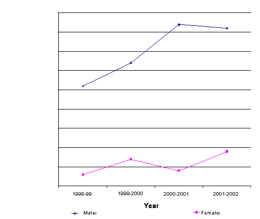
The number of cases increased gradually during the four years period (Figure-1). In facial fractures commonest was the involvement of mandible (62.9%).
Fig-1: Yearly Cases of Maxillofacial trauma
|
0 |
|
5 |
|
10 |
|
15 |
|
20 |
|
25 |
|
30 |
|
35 |
|
40 |
|
45 |
|
1998-99 |
|
1999-2000 |
|
2000-2001 |
|
2001-2002 |
|
Year |
|
Males |
|
Females |
|
No. of Patients |
Majority of the patients in present study had the associated injuries treated concomitantly. These features were not included in some of the other studies carried out in Pakistan6,13. Facial lacerations were closed primarily and patients having the element of head injury were observed and treated by the active participation of the neurosurgery department. Intra oral approach was preferred leaving no residual scar.
Simple method of reduction and immobilization were employed for isolated mandibular fractures for most of the cases whereas other modern modalities of open reduction and internal fixation were also used (which included lag screws, dynamic compression plates, reconstruction plate mini plates, micro plates). Interosseous wiring was also done in 18 cases in early period of study when the other modern modalities of ORIF were not available. Two patients had a lost segment of mandibular body after gunshot injury for which iliac bone graft was harvested and fixed with the dynamic compression plate.
Patients who were managed by only IMF, had a longer duration (4–6 weeks) of immobilization as compared to the patients having ORIF+IMF (3–4 weeks). Far superior results were obtained in only ORIF because of normal mobilization of jaw from the day of surgery and maintenance of weight.
The complications were a few including post-operative pain (7%), malocclusion (4%). In two cases, breakage of reconstruction plate occurred. Plates were palpable in 10.9% of cases. In 12 patients the mental nerve was severed resulting in unilateral lower lip anaesthesia/hypoaesthesia. Ideally we should use the Titenium implants instead of Stainless steel implants. But due to financial constraints of the patients we were bound to use the Stainless steel implants, which resulted in a relatively high complications rate.
Being a developing country, the socio-economic status of the majority is low. Moreover, the patients came to PIMS, being a tertiary care centre, from far flung areas. All these factors contributed to the irregular follow up of the patients. But the available data showed the outcome of the patients was satisfactory and up to the mark.
Most of the accidents occur in the rush hours. The laws regarding the precautions (like seat belts, speed limits, traffic education etc) are not observed. The vehicles are not checked and given proper attention due to obvious socio-economic conditions. All these factors result in increasing the number of road traffic accidents. Now a days in developed countries, to avoid the secondary removal of plates, bioabsorbable plates system has been developed for use in craniomaxillofacial surgery15.
The commonest cause of maxillofacial injuries in our study was road traffic accidents compared to other study8. But the reason of accidents in our country is due to socioeconomic conditions and violation of traffic rules whereas in developed countries, accidents are mostly due to alcoholic intoxication8.
Maxillofacial injuries may cause serious cosmetic and functional deformities. Patients with these injuries are candidates of number of operations. We conclude that early interventions including reduction, stabilization of fractures as well as bone or cartilage grafting (if necessary) will decrease the number of operation and healing period. We also recommend open reduction and internal fixation for maxillofacial fractures should be undertaken whenever possible.
REFERENCES
1. Nayyar MS, Ekanayake MBK. Assessment of maxillofacial injuries. Pakistan Oral Dent J 2001;21:12–8.
2. Muzaffar K. Management of maxillofacial trauma. AFID Dent J 1998;10:18–21.
3. Manson PN. Facial injuries. In: McCarthy JG, editor. Plastic Surgery. Philadelphia: W.B. Saunders;1990:867–1141.
4. Segal Z, Thaller S, Segal A. Facial trauma: zygomatic complex fractures. Emedicine Journal;January 14, 2002. http://www.emedicine.com/face/zygoma.html
5. Obsorn TE, Bays RA. Pathophysiology and management of gunshot wounds to the face. In: Foscla RJ, Walker PV. Oral and maxillofacial trauma. Philadelphia: WB Saunders 1991:672–721.
6. Ambreen A, Shah R. Causes of maxillofacial injuries—a three years study. J Surg Pak 2001;6(4):25–7.
7. Yamaoka M, Furuska K, Fgueshi K. The assessment of fractures of the mandibular condyle by use of computerized tomography: incidence of saggital split fracture. Br J Oral Maxillofac Surg 1994;32:77–9.
8. Gorgu M, Adanali G, Tuneel A, Senen D, Erdogan B. Airbags and wearing seat belts prevent crush injuries or reduce severity of injury in low-speed traffic accidents. Eur J Plast Surg 2002;25:215–18.
9. Gratten E, Hoobs JA. Mechanism of injury to the face in road traffic accidents. In: Rome NL, Williams J, eds. Maxillofacial injury. London: Churchill Livingstone;1985:pp 37–74.
10. Allen MJ, Barens MR, Bodiamala GG. The effect of seat belt legislation on injuries sustained by car occupants. Injury 1985;16:471–3.
11. Shephered JP. Surgical, socio-economic and forensic aspects of assault; a review. Br J Oral Maxillofac Surg 1989;27:89–98.
12. Perkins CS, Layton SA. The aetiology of maxillofacial injuries and the seat belt law. Br J Oral Maxillofac Surg 1988;26:353–63.
13. Zakai MA, Islam T, Memon S, Aleem A. Pattern of maxillofacial injuries received at Abbasi Shaheed Hospital, KMDC, Karachi. Annals Abbasi Shaheed Hosp 2002;7:291–3.
14. Ugboko VI, Obusanuya SA, Iagade OO. Maxillofacial fractures in a semi urban Nigerain teaching hospital. Int J Oral Maxillofac Surg 1998;27:286–9.
15. Zachariades N, Papavasilliou D, Papadimetrio I, Koundouris T. Fracture of the facial skeleton in Greece: a retrospective study covering 1791 cases in 10 years. J Maxillofac Surg 1983;11:142–4.
16. Kim YK, Kim SG. Treatement of mandiibular fractures using bioabsorbable plates. Plast Reconstr Surg 2002;110:25–31.
Address for correspondence:
Dr. Syed Shahid Hussain, Resident, Plastic Surgery, Room No. 34, M.O. Hostel, P.I.M.S., Islamabad, 44000, Pakistan.
E-mail: shahid_pims@hotmail.com