INTRODUCTION
Central nervous system (CNS) infections are a group of life threatening diseases that present a formidable challenge to physicians. Despite the development of effective antibiotics and modern surgical techniques significant mortality and morbidity persist among patients with CNS infections. Since the introduction of cross sectional imaging earlier diagnosis has become possible and this has led to a decrease in both morbidity and mortality. The use of CT has brought a marked disease in mortality among patients with brain abscesses1 and the advent of magnetic resonance imaging (MRI) has accelerated this trend. Currently, Mri has become the modality of choice in the evaluation of suspected CNS infection but it is at least four time expensive investigation and is available in very few hospitals. So in our set up CT Scan is a useful investigation in the early diagnosis of CNS infection.
Infections reach the brain or meninges predominantly by two routes: (a) hematogenous dissemination from a distant infective focus to the meninges, corticomedullary junction and choroid plexus and (b) direct extension from an adjacent focus of suppuration (otitis, mastoiditis, sinusitis) or a traumatic craniocerebral wound.
MATERIALS AND METHODS
The study included 250 consecutive patients referred to the radiology department of Ayub Teaching Hospital for CT scan brain with the complaints of high grade fever and:
(a) headache/neurological symptoms
(b) otitis/mastoiditis or
(c) sinusitis.
Transaxial 5/10mm sections were obtained pre I/V contrast and 3/5/10mm sections were obtained post I/V contrast according to size of the lesion. 5mm coronal sections were also obtained where source of infection was suspected to be in the sinuses/petrous bone and in some cases for better visualization of the lesions. Follow-up CT scans were performed in a few cases.
RESULTS
Among the different CNS infections in our study, meningitis has the highest frequency 21.6% followed by abscesses. Encephalitis and tuberculomas are also fairly common. A few cases of sub-dural empyema and a single case of hydatid cyst were also found.
Table-1 shows the CT diagnosis of brain infections in 250 symptomatic patients. Fig-1 shows the age distribution of brain infection and Fig-2 shows the gender distribution of brain infections.
Table-1: CT Diagnosis of brain infections in 250 symptomatic patients
|
|
No. of Patients |
Percentage |
|
Meningitis |
54 |
21.6 |
|
Brain abscess |
31 |
12.4 |
|
Encephalitis |
15 |
6.0 |
|
Tuberculomas |
9 |
3.6 |
|
Sub dural empyema |
4 |
1.6 |
|
Hydatid cyst |
1 |
0.4 |
|
Normal |
136 |
54.4 |
|
Total |
250 |
100% |
Fig. 1: Age Distribution of brain infections
Figure-2: Gender Distribution of brain infections.
DISCUSSION
CNS infections are quite common especially at the two extremes of age probably due to reduced immunity. In our study of 114 positive cases, 55.26% patients were under 20 years and 22% were above 60 years. So 77% cases were at the two extremes.
Uncomplicated meningitis is usually silent in the computed tomogram. However, in fulminant cases meningeal and cortical enhancement is seen following contrast medium administration2. CT is also very useful in the detection of complications of meningitis such as thrombosis of the draining venous sinuses, infarction, hydrocephalus and subdural effusions which may develop into subdural empyemas3.(Fig: 3,4)

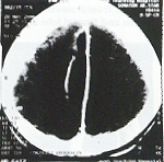
Figure-3: Right dided inter-hemisphere sub-dural empyema delineated by thick falx cerebri medially and an early membrane formation on the lateral aspect.
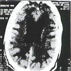
Figure-4: Dilated ventricles with CSF-fluid/pus level-ventriculitis.
Cerebral abscess is an encapsulated inflammation. It is easily diagnosed or excluded by CT scan. The CT appearance of an abscess is that of a well-defined hypodense area showing ring enhancement and accompanied by extensive perifocal oedema and mass effect. In about 50% cases the medial wall of an abscess is thinner than the lateral one and is thought to be due to the relatively poor vascular supply of the white matter. This explains the tendency of abscesses to rupture into the ventricles and the development of secondary abscesses medially4. Smooth less than 5mm thick wall with medial thinning helps in differentiating an abscess from a cystic tumour. (fig. 5,6)

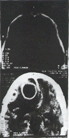
Figure-5: Right frontal sinusitis causing osteomyelitis and frontal lobe abscess
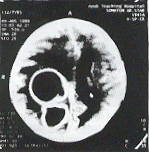
Figure-6: Two abscesses in the right parietal lobe
Most of the encephalitides resemble each other and have few identifying imaging characteristics. Areas of involvement show oedema, mass effect and perhaps petechial haemorrhages. A few encephalitides have typical features like herpes simplex type I encephalitis, in which the most commonly noted CT findings are poorly marginated hypodense areas with mass effect and nonhomogenous contrast enhancement in the temporal and frontal lobes5,6. Similarly varicella encephalitis may particularly affect the cerebellum, even when pan encephalitis is seen7. Therefore the primary role of CT scan in the evaluation of encephalitis is to support the clinical diagnosis, indicate the best site for biopsy and to exclude brain abscess or tumour. (Fig 7)
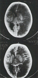
Figure-7: Hypodense oedematous swelling of left hemisphere with cortical hyperaemia-Encephalitis
The most common cause of subdural empyema is para nasal sinusitis8. Before the era of CT, subdural empyema was associated with a mortality rate as high as 40%. Since the advent of CT the mortality has dropped significantly9,10.
Cerebral tuberculomas although rare in the West are quite common in this part of the world. The clinical features of tuberculomas are rarely distinguishable from those of other space occupying lesions11 and 42% of patients with intacranial tuberculomas have no evidence of extra carnial disease12. So CT scan helps in the correct diagnosis of this easily treatable condition. (Fig. 8)
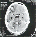
Figure-8: Multiple rounded enhancing lesions with perifocal oedema with mass effect-Tuberculomas
Figure-9: Hydatid cyst left parietal lobe
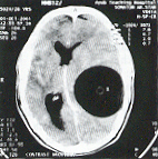
Hydatid disease is usually manifested by cysts in the liver and lungs. Only 2% of hydatid infections involve the brain of man and is always correctly diagnosed by CT. We had a single case of hydatid cyst in the parietal lobe. It is usually large, spherical and solitary. The CSF like density of the cyst contents and the absence of perifocal and peripheral contrast enhancement differentiate this lesion from cerebral abscess on CT13. (Fig.9)
CONCLUSION
The study has strongly supported the importance of CT scan in the early and correct diagnosis of brain infections.
REFERENCES
1. Roienbaum ML. Decreased mortality from brain abscesses since the advent of CT. J Neuro Surg 1978;49:659-668.
2. Moseley JF. The role of CAT in diagnosis and management of intracranial infections In: Computed axial tomography Berlin: Springer 1977.
3. Claveria LE, Duboulay GH, Moseley JF. Intra cranial infections: Investigation by CAT. Neuroradiology 1976; 12:59-71.
4. Stevens EA. CT brain scanning in intraparenchymal pyogenic abscesses. AJR 1978; 130:111-114.
5. Davis JM. CT of herpes simplex encephalitis with clinicopathological correlation. Radiology 1978;129:409-417.
6. Leo JS. CT in herpes simplex encephalitis. Surg Neurol 1978;10:313-317.
7. Hurst DL, Mehtu S. Acute cerebellar swelling in varicella encephalitis. Pediatr Neurol 1988; 4:122-123.
8. Carter BL, Bankof MS, Fisk JD. CT detection of sinusitis responsible for intracranial and extra cranial infections. Radiology 1983;147:739-742.
9. Weisberg L. Subdural empyema clinical and computed tomograph relations. Arch Neurol 1986;43:497-500.
10. Weinman D, Samarashinghe HHG. Subdural empyema. Aust NZJ Surg 1972;41:324-328.
11. Anderson JM, Macmillan JJ. Intracranial tuberculoma. An increasing problem in Britain. J Neurol Neurosurg Psychiatry 1975;38:194-201.
12. Mayers MM, Kaufman DF, Miller MM. Recent cases of Intracranial tuberculomas. Neurology 1978;28:256-260.
13. Abbassioun K. CT in hydatid cyst of the brain. J Neurosurg 1978;49:408-411.
