INTRODUCTION
Evaluation
of a female patient who presents with acute abdomen always remains a challenge
and it becomes more troublesome when a pregnant patient presents with acute
abdomen1. Non-obstetric surgery in the pregnant patient can be both
diagnostically and technically challenging2. Diagnosis of
appendicitis in pregnancy is difficult, as in other abdominal surgical
conditions. The symptoms are non-specific and most often are attributed to
pregnancy itself1-3. Diagnostic delays tend to occur in pregnant
patients for many reasons: first, and the most important is the
misinterpretation of signs and symptoms of acute appendicitis with the
pregnancy, both by the patient and her treating physician. Second, the
pregnant abdomen is difficult to examine and usually hide or change the
classical signs of acute appendicitis and lastly many of physicians are more
conservative with pregnant patients and this may actually tend to do more harm
by causing a delay in diagnosis and treatment1,4,5. It is well
known that the delay in diagnosis and definitive treatment represents the most
significant risk for poor outcome on both mother and her foetus6.
In 1908 it was first reported that the mortality of appendicitis complicating
pregnancy is the mortality of delay7. This holds true for any
condition that would cause an acute abdomen in pregnancy: however, surgical
diseases in pregnancy are a rare event, there remains a lack of data on the
indications for operation, approach of operation, and risk to mother and
foetus8. This present study describes our experience with the
management of acute appendicitis complicating pregnancy.
MATERIALS
AND METHODS
This study was carried out at Department of surgery; East Surgical Unit, Mayo Hospital Lahore, from January 1999 to July 2001. It included a total of 50 pregnant patients who presented to the emergency department with probable diagnosis of acute appendicitis. All the patients were admitted, after resuscitation full history and thorough clinical examination were recorded. The history included the site of pain, its onset, character, migration, radiation, aggravating and relieving factors and any associated symptoms like nausea, vomiting, fever etc. The period of gestation was noted. Any problems and or complication during previous pregnancies were noted. The clinical examination included general physical examination, abdominal examination and pelvic examination. Laboratory investigations included Total Leukocyte Count and urine examination. Abdominal ultrasonography was performed where delay in the surgical treatment could be tolerated both by the patient and her treating surgeon. Prompt surgical intervention was done when the diagnosis of acute appendicitis was established. In patients where the doubt existed, serial examinations were performed to confirm the diagnosis. Third generation Cephalosporin in dose of 1 gram I/V was administrated during induction of Anaesthesia and post operatively b.i.d. for three days. Foetal heart sounds were monitored postoperatively to ensure foetal well-being; in cases of any doubt foetal ultrasound was performed on second postoperative day. Patients were followed up for their disease and out come of the pregnancy, including maternal and foetal mortality and morbidity.
RESULTS
Over
a period of thirty months, 50 patients were selected for this prospective
study, where follow up was possible in postoperative period. The mean age was
26.5 years (range: 19–36 years) (Table-1).
Table-1: Age frequency
|
Age |
No. of patients |
Percentage |
|
19-25 |
7 |
14 |
|
26-30 |
34 |
68 |
|
31-36 |
9 |
18 |
Out
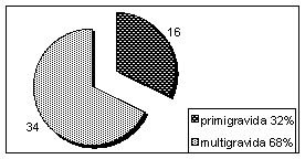
of these 50 patients, 16 were primi-gravida and 34 were multi-gravida, with no
history of acute abdomen during previous pregnancies (Figure-1).
Figure-1: Parity of patients:

Most of these patients were in their second trimester (n= 26), followed by first trimester (n= 19) and third trimester (n= 5). Table-2.
Table-2:
Duration of pregnancy
|
Trimester |
No. of Patients |
Percentage |
|
First |
19 |
38 |
|
Second |
26 |
52 |
|
Third |
5 |
10 |
|
Total |
50 |
100 |
All
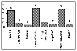
the patients had history of pain abdomen, pain right iliac fossa was observed
in 36 (72%) patients, vague generalized abdominal pain was encountered in 9
(18%) patients and backache was observed in 5 (10%) patients. 41 patients had
history of nausea and vomiting, 11 had associated burning micturation, 7 had
temperature of more than 99 ºF. Thirty-nine patients had WBC count of more
than 15,000/cmm and 7 patients had more than 20 pus cells in urine examination
(Figure-2).
All
the patients under went laparotomy; in 43 (86%) patients the operative
findings supported the clinical diagnosis of acute appendicitis whereas in 7
(14%) patients the results of the laparotomy were negative (Table-3).
Table-3: Results of Surgery
|
Results
|
No. of Patients |
Percentage |
|
Positive
laparotomy |
43 |
86 |
|
Negative
laparotomy |
7 |
14 |
|
Total
|
50 |
100 |
Figure-2: Presentations

All the patients were followed up for the out come of surgery in terms of symptomatology and out come of pregnancy in terms of preterm labour and maternal and foetal mortality and or morbidity. Four of these patients had preterm labour, two weeks before the expected date and all were in their third trimester at the time of surgery. There was no mortality and or morbidity noted in mother during labour, unfortunately the foetal mortality rate was 14% (n= 7) and foetal morbidity was noted in 4 cases with the babies having low birth weight (Table-4).
Table-4:
Outcome of Surgery
|
Outcome |
No.
of Cases |
Percentage |
|
Maternal
mortality |
0 |
0 |
|
Maternal
morbidity |
4
(preterm labour) |
8 |
|
Foetal
mortality |
7 |
14 |
|
Foetal
morbidity |
4
(low birth weight) |
8 |
DISCUSSION
Acute
appendicitis is the commonest non-gynaecological surgical problem occurring
during pregnancy9, with an estimated frequency of one case of acute
appendicitis per 1500 pregnancies10. The incidence of appendicitis
is unchanged in pregnancy, but the clinical presentation becomes even more
variable11. During pregnancy the appendix migrates in a
counter-clockwise direction toward the right kidney, rising above the iliac
crest at about 4.5 months gestation. Right lower quadrant pain and tenderness
dominate in the first trimester, but in the latter half of pregnancy, Right
Upper Quadrant (RUQ) or right flank pain must be looked upon as a possible
sign of appendiceal inflammation. Nausea, vomiting, and anorexia are common in
uncomplicated first trimester pregnancies, but their reappearance later in
gestation should be viewed with suspicion12. The studies of Baer et
al4 in 1932 are well known, showing the migration of the
appendix progressively upwards in right lower and upper quadrants through the
pregnancy. This migration shifts the point of maximum tenderness and also
obscures the classical sign of rebound tenderness13. The WBC counts
increases normally during pregnancy and can reach levels of 16,000/cmm,
therefore, a leukocytosis must be interpreted carefully14.
Acute appendicitis can occur at any point
during gestation but is most common in the first and second trimesters6.
According to a study conducted at Saudi Arabia15, there were 10
(19%) patients who presented in the first trimester, 31 (60%) second
trimester, 8 (15%) third trimester and 3 (6%) patients in the puerperium. Our
results match with these results since most of the patients presented in
second trimester 52% (n= 26), followed first trimester in 38% (n= 19) and
third trimester 10% (n= 5). Due
to difficulty in clinically diagnosing acute appendicitis, the negative
laparotomy rate is much higher in the pregnant than the non-pregnant patients16,17.
An accepted rate of normal appendices in non-pregnant patients undergoing
laparotomy for suspected appendicitis is 15%. This has been much higher in
pregnant patients, with larger series having a misdiagnosis rate between
approximately 20% and 35%10. Similarly according to the study by
Masters et al16, the rate
of positive laparotomy was 81% and that of negative was 19%.
Our present study correlates with all these
studies as we have 86% positive laparotomy and 14% negative laparotomy. It
may, however, be important to have a higher negative laparotomy rate in
pregnant patient with suspected appendicitis secondary to the grave
consequences of missing the diagnosis. The foetal mortality increases
dramatically if perforation occurs or appendicluar abscess develops. Foetal
loss occurs in 3% to 5% of cases of acute appendicitis but increases to 20%
with perforation and abscess18. An aggressive surgical approach is
therefore justified. In two separate large institutional reviews,
non-obstetric intra-abdominal surgery was reported to have a frequency of 1in
451 to 1 in 635 deliveries2,8. Both series confirmed that intra
abdominal surgery during pregnancy carries an acceptable risk to both the
mother and the foetus and that complications are related to disease severity
and operative delay rather than the operative procedure itself2.
Overall risk of preterm labour has been reported to be between 4% to 6% with
pelvic or lower abdominal surgery19,20, others have reported this
risk to be 15% to 20%2 even up to 38%8, our study shows
preterm labour in 8% of patients and foetal mortality of 14 %, which obviously
correlates with international and local21 series.
CONCLUSION
From
this study, we conclude that: (1) misdiagnosis of appendicitis in pregnancy is
comparable to that in the general female population;
(2) foetal mortality is minimal with early operation before perforation
(3) clinical judgment rather than laboratory remains the gold standard for the
diagnosis; and (4) the pregnant patient presenting with abdominal pain should
be assessed and treated as one would any patient with the same complaint,
i.e., unnecessary delay should be avoided as it is directly related to
maternal and foetal morbidity and mortality. The general use of this principle
may explain the marked improvement in maternal and foetal mortality and
morbidity in recent years.
REFERENCES
1.
Hector MT, Robert DM. Gynecological causes of the acute abdomen and the
acute abdomen of pregnancy. Surg Clin North Am 1997;77 (6):1371-1393.
2.
Allen JR, Helling TS, Langerfeld M. Intra-abdominal surgery during
pregnancy: Am J Surg 1989;158:567.
3.
Kesarwani RC. Acute appendicitis complicating pregnancy: J Indian Med
Assoc 1984;82(9): 316-8.
4.
Baer JL, Reis RA, Aren RA: Appendicitis in pregnancy with change in
position and axis of the normal appendix in pregnancy. JAMA 1932; 98:1359.
5.
Bello GV, Schonholz D, Moshirpur J. Comboned pregnancy: the Mount Sinai
experience. Obstet Gynecol Surv 1986; 41:603.
6.
Fallon WF, Newman JS, Fallon GF. The surgical management of intra
abdominal inflammatory conditions during pregnancy: Surg Clin North Am 1995;
75:15.
7.
Bable EA. Perforated appendicitis complicating pregnancy. JAMA
1908;51:1310.
8.
Kort B, Katz VL, Watson WJ. Effect of non obstetrical operation during
pregnancy. Surg Gynecol Obstet 1993; 177:37.
9.
McGee TM. Acute appendicitis in pregnancy. Aust N Z J Obstet Gynaecol
1989;29(4): 378-85.
10.
Mazze RI, Kallen B. Appendectomy during pregnancy: A Swedish Registry
study of 778 cases. Obstet Gynecol 1991;77:836.
11.
McGee TM. Acute appendicitis in pregnancy: Aust N Z J Obstet Gynaecol
1989;29(4):378-85.
12.
Sandy C, William L, Francisco T, Eugene H: Acute Appendicitis.
eMedicine Journal 2001; 2(7).
13.
Cunningham FG, McCubbin JH. Appendicitis complicating pregnancy. Obstet
Gynecol 1977;45:415.
14.
Choudhary S, Andley R, Sharma VP, Bhatnagar R. Assessment of leucocytes
count in the diagnosis of acute appendicitis. J Indian Med Assoc
1980;74(5):85-7.
15.
Al-Mulhim AA. Acute appendicitis in pregnancy: A review of 52 cases.
Int Surg 1996;81(3):295-7.
16.
Masters K, Levine BA, Gaskill HV, Sirinek KR. Diagnosing appendicitis
during pregnancy: Am J Surg. 1984;148(6):768-71.
17.
Bailey LE, Finley RK Jr, Miller SF, Jones LM. Acute appendicitis during
pregnancy: Am J Surg. 1986;52(4):218-21.
18.
Mahmoodian S. Appendicitis complicating pregnancy: South Med J
1992;85:19.
19.
Hunt M G, Martin JN Jr, Martin RW. Preinatal aspects of abdominal
surgery for non-obstetrical diseases: Am J Perinatol 1989; 6:412.
20.
Kammerer W D: Non-obstetrical surgery in pregnancy: Med Clin North Am
1987; 71:551.
21.
Syed S. Difficulties in diagnosis of acute appendicitis in pregnancy.
J Pak Med Assoc 1985;35(9):282-5.
Address for
Correspondence and Reprints:
Dr.
Abbas Ghazanfar. East Surgical Unit. Mayo Hospital Lahore. Pakistan.